Deep Vein Thrombosis (DVT)
Definition | Aetiology | Pathophysiology | Risk Factors | Signs and Symptoms | Investigations | Management | Patient Advice
Definition
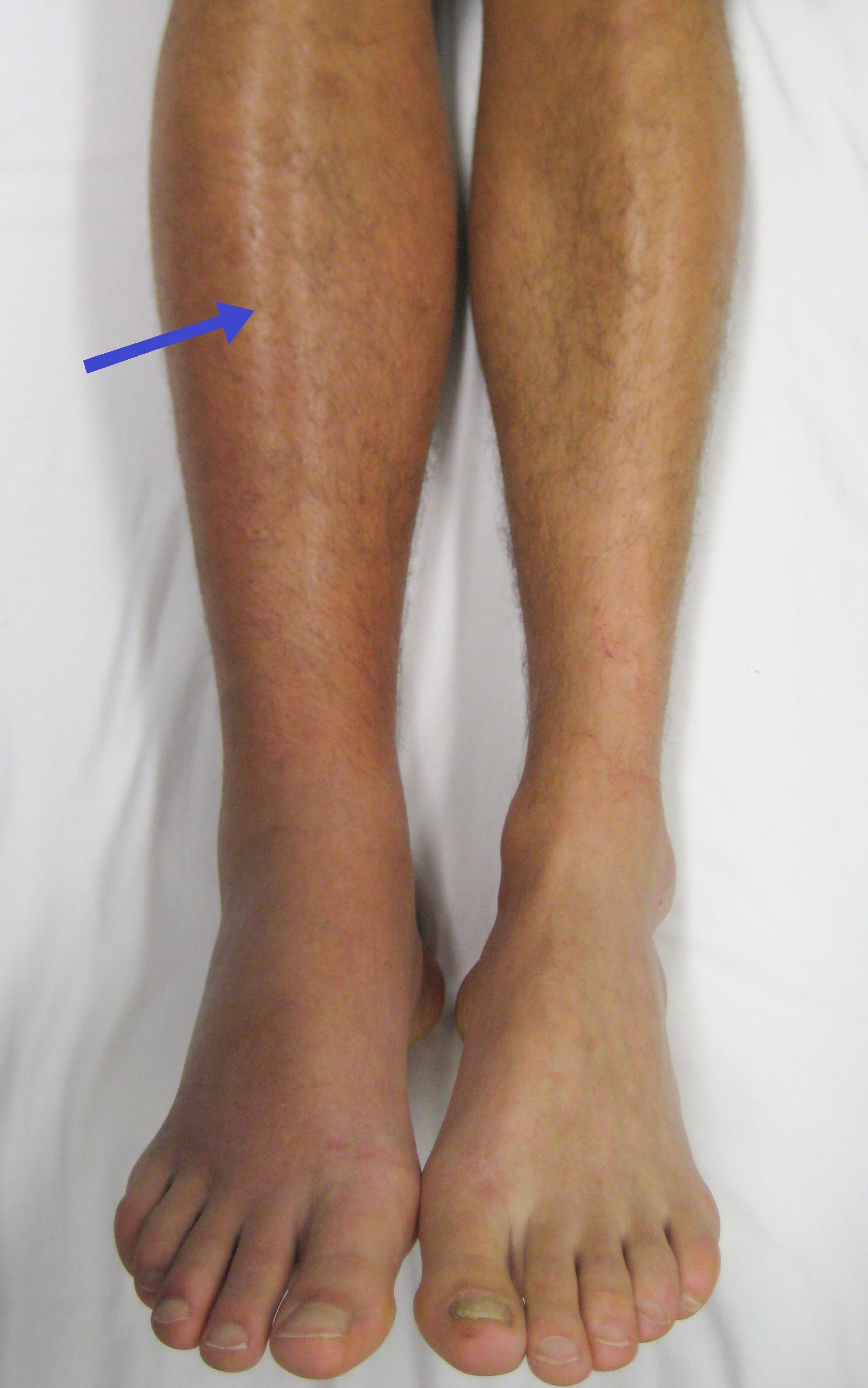
Deep Vein Thrombosis (DVT) is the formation of a blood clot in a deep vein, most commonly in the legs. It can cause pain and swelling and carries a risk of life-threatening complications such as pulmonary embolism (PE). See Figure 1.
Aetiology
DVT results from a combination of factors that contribute to clot formation, known as Virchow's triad:
- Venous Stasis: Reduced blood flow in veins due to immobility, prolonged bed rest, or long-haul flights.
- Endothelial Injury: Damage to the vein wall from trauma, surgery, or inflammation.
- Hypercoagulability: Increased clotting tendency due to conditions such as pregnancy, cancer, or inherited thrombophilias (e.g., Factor V Leiden).
Pathophysiology
DVT develops due to the following processes:
- Clot Formation: Platelets and fibrin aggregate to form a thrombus within the vein.
- Venous Obstruction: The clot obstructs blood flow, leading to swelling and increased venous pressure.
- Risk of Embolisation: The clot may dislodge and travel to the lungs, causing pulmonary embolism.
Risk Factors
Key risk factors include:
- Prolonged immobility (e.g., after surgery or long travel).
- Recent surgery, particularly orthopaedic or pelvic procedures.
- Pregnancy or postpartum period.
- Cancer or chemotherapy.
- Use of oestrogen-containing contraceptives or hormone replacement therapy.
- Obesity or smoking.
- Inherited thrombophilias (e.g., Factor V Leiden mutation).
Signs and Symptoms
Symptoms of DVT include:
- Swelling: Unilateral swelling of the affected leg.
- Pain: Calf or thigh pain, often described as cramping or aching.
- Redness and Warmth: Over the affected area.
- Homan's Sign: Pain in the calf on dorsiflexion of the foot (not specific).
Investigations
Key investigations and common positive findings include:
- Wells Score: A clinical scoring system to assess DVT risk.
- D-dimer Test: Elevated levels indicate active clot formation and breakdown, though not specific for DVT.
- Compression Ultrasound: The diagnostic test of choice. Positive findings include non-compressibility of the affected vein.
- Venography: Used in complex cases to visualise the clot directly, though rarely needed.
Management
1. Primary Care Management
- Immediate Referral: Urgent referral to secondary care if DVT is suspected.
- Initial Anticoagulation: If available, administer low-molecular-weight heparin (e.g., enoxaparin) while awaiting secondary care review.
2. Secondary Care Management
- Anticoagulation Therapy: Start with low-molecular-weight heparin, transitioning to oral anticoagulants such as apixaban or rivaroxaban.
- Monitoring: Regular INR monitoring if using warfarin.
- Thrombolysis: Considered in massive DVT or phlegmasia cerulea dolens, typically performed by an interventional radiologist.
- Inferior Vena Cava (IVC) Filter: For patients with contraindications to anticoagulation, performed by a vascular specialist.
Patient Advice
Key advice includes:
- Take anticoagulants as prescribed and attend regular follow-ups.
- Stay mobile and avoid prolonged immobility.
- Maintain a healthy weight and stop smoking.
- Wear compression stockings to prevent post-thrombotic syndrome, if recommended.
- Seek immediate medical attention for symptoms of pulmonary embolism, such as sudden chest pain or breathlessness.
Figure 1

Image showing a deep vein thrombosis in the right leg, with swelling and redness.
References
- James Heilman, MD (2015). Deep Vein Thrombosis of the Right Leg [Image]. Available at: https://upload.wikimedia.org/wikipedia/commons/2/21/Deep_vein_thrombosis_of_the_right_leg.jpg (Accessed: 30 December 2024).
| Clinical Feature | Points |
|---|---|
| Active cancer | 1 |
| Paralysis, paresis, or immobilization of lower extremity | 1 |
| Recently bedridden for more than 3 days, or major surgery within the past 12 weeks | 1 |
| Localized tenderness along the distribution of the deep venous system | 1 |
| Entire leg swelling | 1 |
| Calf swelling at least 3 cm larger than asymptomatic side (measured 10 cm below tibial tuberosity) | 1 |
| Pitting edema confined to symptomatic leg | 1 |
| Collateral non-varicose veins | 1 |
| Previously diagnosed DVT or PE | 1 |
Interpretation:
A score of 0 or less: low probability of DVT
A score of 1-2: moderate probability of DVT
A score of 3 or more: high probability of DVT
If suspected DVT refer to local DVT clinic
Check out our youtube channel
Blueprint Page
Explore the comprehensive blueprint for Physician Associates, covering all essential topics and resources.
Book Your Session
Enhance your skills with personalised tutoring sessions tailored for Physician Associates.

